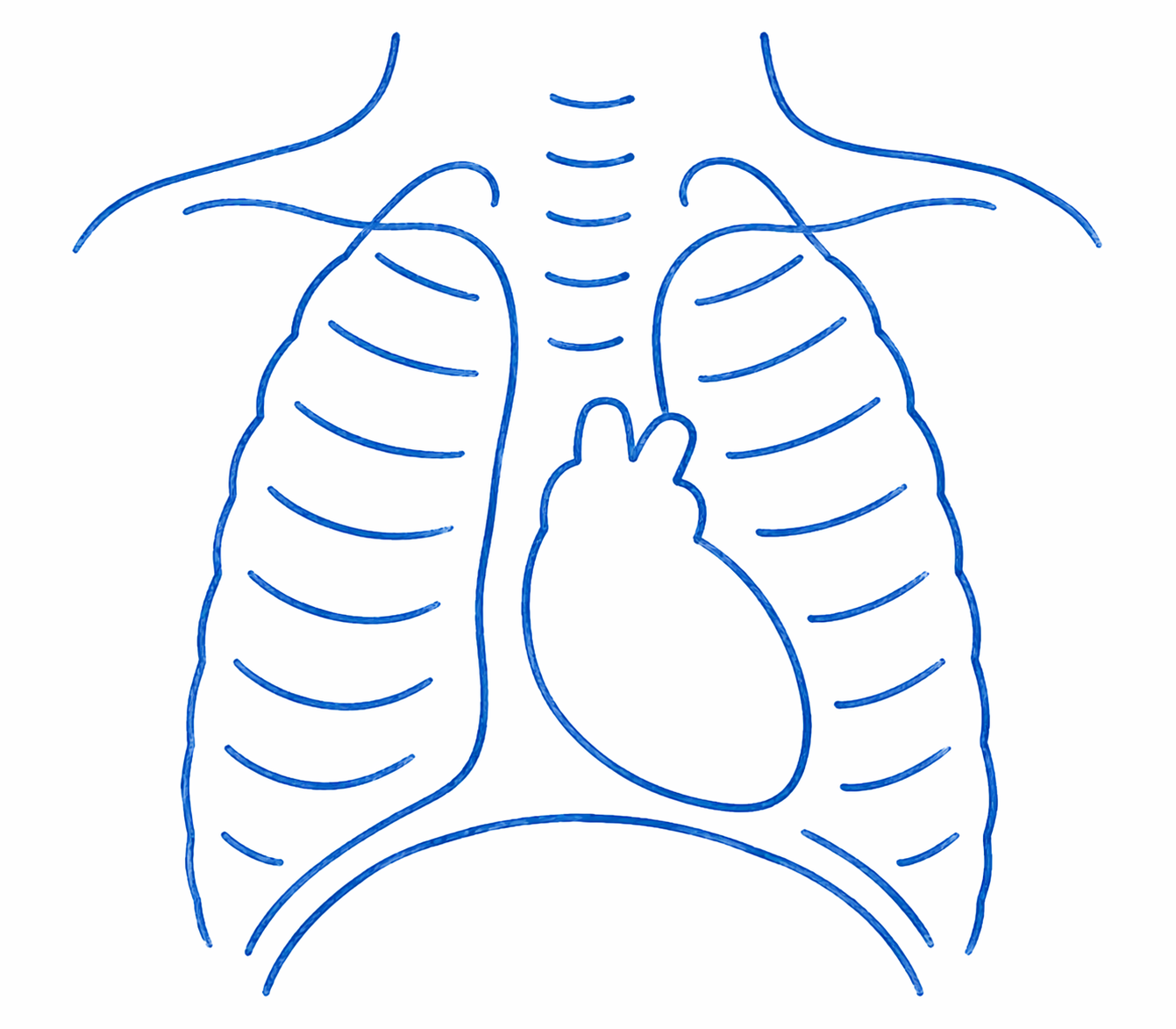
A quick, painless picture of the heart and lungs using a very low dose of radiation. Shows heart size, lung blood flow and airway health. Takes only seconds and needs no special preparation.
Electrophysiologic (EP) Study and Ablation
A procedure to find and treat abnormal heart rhythms. Thin catheters are guided to the heart to map electrical signals and, if needed, ablation uses heat or cold to permanently correct the problem. Performed under general anaesthetic.
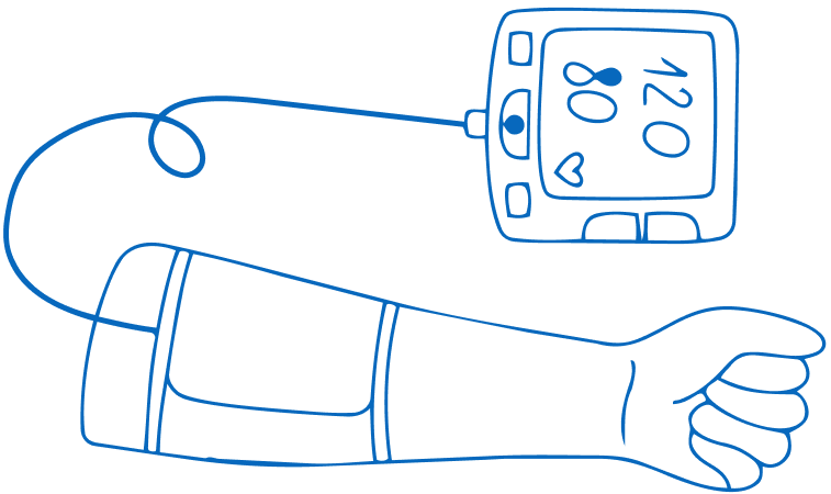
Blood pressure monitoring
Measures the force of blood in the arteries using an arm cuff. A 24-hour ambulatory monitor can be worn to record blood pressure during daily activities and sleep, giving a complete picture. Quick, safe and painless.
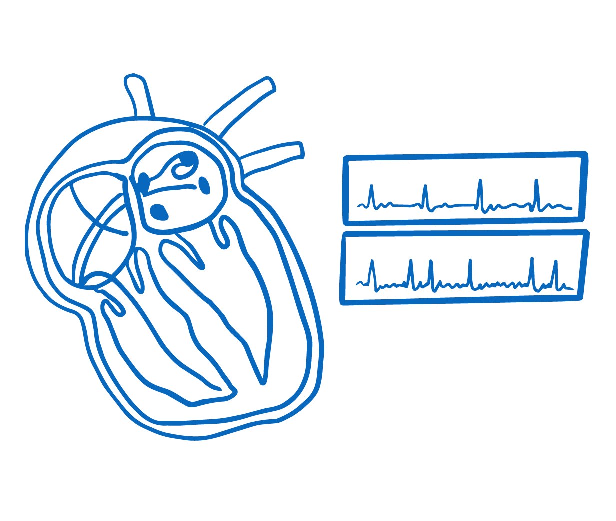
Rhythm monitoring/Holter ECG
Extended recording of the heart rhythm over hours or days to catch abnormal beats. Options include a 24-hour Holter monitor and a wireless patch worn for up to 2 weeks.
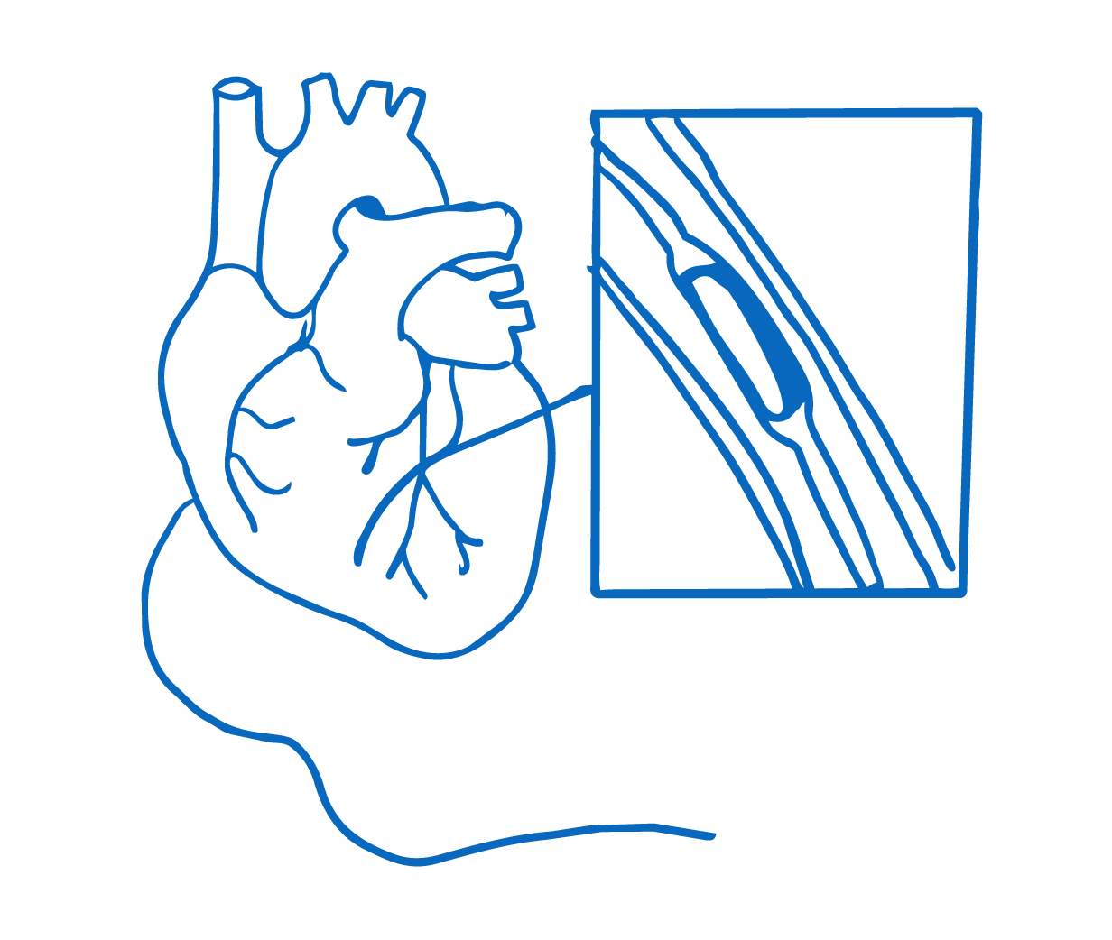
Cardiac catheterization and Keyhole procedures
A keyhole procedure using a thin tube guided through a blood vessel to the heart. Measures pressures, takes X-ray images and can treat many conditions without open surgery. Performed under general anaesthetic.
Advanced scans producing detailed images of the heart and blood vessels. MRI uses magnets and radio waves with no radiation; CT uses X-rays with a very low dose. Both need a small cannula for contrast dye. Safe and highly accurate.
Consists of the controlled measurement of a child heart, lungs, circulation and muscles responses to exercise. Can investigate the causes of chest pain, and palpitations, of fainting during exercise, of breathlessness during exercise, including asthma and of easy fatigue.
A safe, painless ultrasound scan of the heart. A small probe with warm gel is placed on the chest to create detailed images of heart chambers, valves and blood flow. No needles or radiation. Results are usually available straight away.
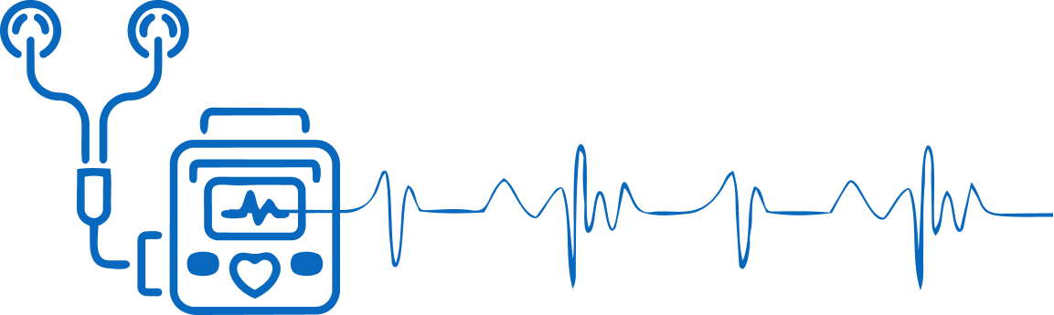
The ECG consists in the recording of the electrical activity of the heart on paper. ECG is performed by applying 10 small stickers to the child skin, 6 on the chest and one each in each arm and leg. The stickers have the purpose of picking up the heart’s electrical signals.
The examination consists in the doctor examining the chest and listening to the chest with a little microphone called stethoscope to try and spot some clues which might be able to establish the cause of the symptom or complaint. This is the way most heart murmurs are found.


.png)